Contact
Lab Head
Postal address
Methods
Neuroanatomy / Microscopy
Immunohistochemistry: double- and triple-labelling with differently coloured fluorescent markers, confocal microscopy (right example)
Immunohistochemistry: DAB-labelling
In-situ hybridisation
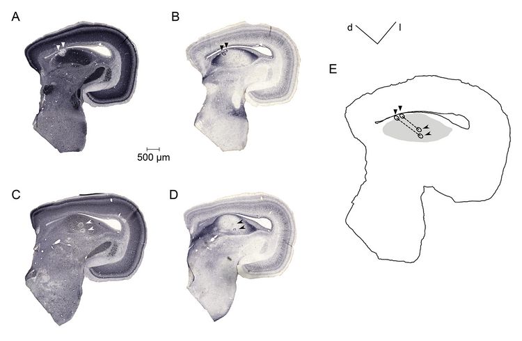
Dye or lesion marking of recording sites (left example)
Electrophysiology in-vivo
Recording of evoked potentials with, e.g., subcutaneous metal electrodes
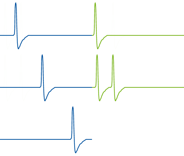
Recording of single-unit action potentials with glass microelectrodes
Molecular Genetics
RT-PCR on developmental cDNA libraries from chicken embryos
Cloning of expression vectors
Production of retrovirus stocks in cell culture
Virus-mediated long-term and stable gene misexpression in chicken embryos
Design and application of short hairpin (sh)RNA interference
Optogenetics
Expression of light-activated proteins (green label in the image) in neurones of the barn owl's brain. The foreign gene was introduced by a virus, injected into the brain some weeks earlier.
We are currently optimizing the light stimulation, via a fine glass fibre (gray in the image) that is connected to a laser at the other end. Neural acitivity is simultaneously registered by an electrode (black).
Stay tuned .....