Single crystal X-ray diffraction
Downloads
Single crystal X-ray diffraction
Latest
Lukas Bührmann, Amrit Chandi, Nadeschda Geibel, Lena Albers, Marc Schmidtmann, Thomas Müller, Boragerma[5]pyramidanes via a Germole-to-Borole Rearrangement, Inorg. Chem. 2026, 65, 7322−7332
Instruments
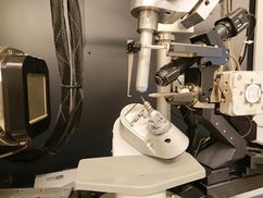
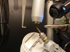
Bruker AXS D8 Venture
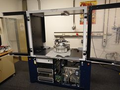
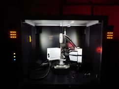
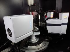
- IµS 3.0 molybdemum microfocus source, multilayer optics
- IµS 3.0 copper microfocus source, multilayer optics
- Photon III CPAD detector
- Oxford Cryostream 800+ (80-500 K)
Bruker AXS Apex II CCD
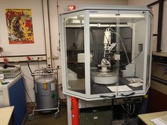
- Molybdenum sealed tube, graphite monochromator
- Apex II CCD detector
- Kryoflex sample cooling (90-300 K)
Method
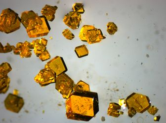

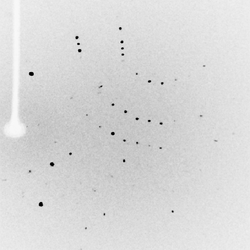
Single crystal X-ray diffraction is a tool to determine the 3-dimensional structure of matter on an atomic level. What you need is single crystals and X-rays, where the crystal is exposed to the X-rays. A crystal is highly symmetrical and ordered which, because of interference effects, results in diffraction of the X-rays. The diffracted X-rays are detected in form of spots (reflections) by a detector. Analysis of the position and intensities of these reflections allows the determination of the 3-dimensional structure of atomic or molecular compounds. XYZ coordinates are obtained for each atom which can be used to calculate bond lengths and angles amongst other geometrical parameters, all important for the chemist.
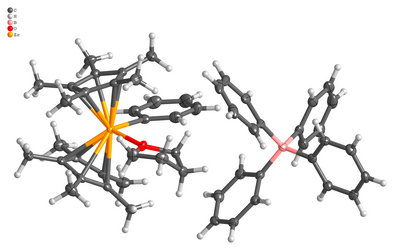
The result of the single crystal X-ray experiment is the 3-dimensional atomic or molecular structure of a compound. The compound fills up the space in the unit cell (black box in below video) which is the smallest building block to build up the visible crystal only by translation in the 3 dimensions. A typical unit cell content is shown below.