


The activity of nerve cells in the brain generates extremely weak magnetic fields. Oldenburg psychologists are using sophisticated techniques to measure these brainwaves and gain further insights into the workings of the brain.
Neuroscientists go to great lengths to record the magnetic signals of a human brain: the body and clothes of test persons must be absolutely free of metals. Even glittery nail polish can distort measurements, as would a train passing by several hundred metres away, not to mention electronic devices. “The magnetic signals generated by brain activity are in the femtotesla frequency range, so more than a billion times weaker than the Earth’s already weak magnetic field,” explains Dr. Florian Kasten, a postdoctoral researcher in the General Psychology research group.
Kasten is studying these barely measurable signals to gain a better understanding of how the human brain works. Together with Prof. Dr. Christoph Herrmann, head of the research group, and other colleagues, Kasten recently achieved a breakthrough in improving understanding of the complicated effects of non-invasive brain stimulation that result from individual variations in neuroanatomy.
Tiny magnetic oscillations
The key tool for such investigations is a device that can detect the tiny magnetic oscillations using a technique called Magnetoencephalography (MEG). “This technique provides similar data to electroencephalography, measuring the activity of large groups of nerve cells in the brain, but it is more effective for pinpointing the sources of the activity,” Kasten explains. Researchers trying to find out where in the brain certain processes take place therefore use MEG measurements, generally in combination with magnetic resonance imaging (MRI), which produces high-resolution images of anatomical structures.
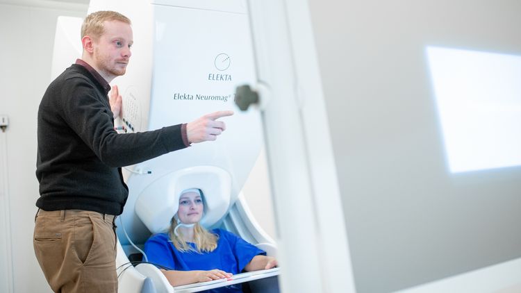
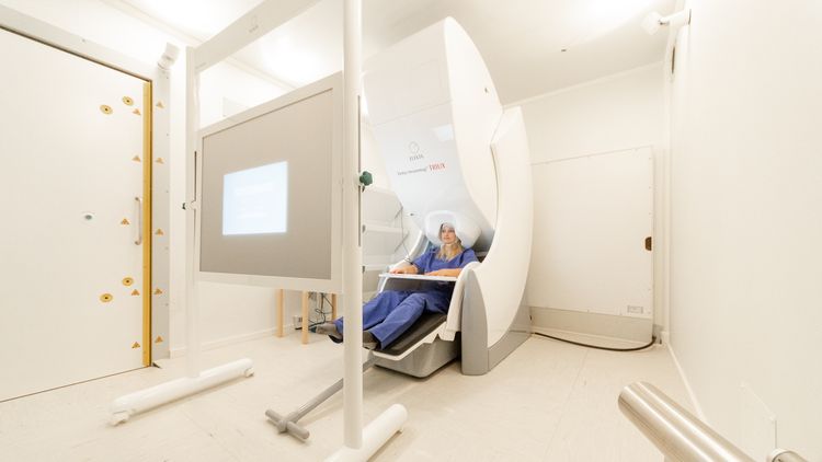
For some time now the University has owned a magnetoencephalography scanner that cost approximately three million euros – one of only around ten such devices in Germany. The white colossus is housed in a special room in the NeSSy research centre on the Wechloy Campus. The room, known as a “measuring cabin”, rests on special foundations that absorb vibrations. It is locked behind a hydraulic door half a metre thick and covered in a special layer of metal that blocks most external magnetic fields. “These measures ensure that we have a very stable magnetic field in the cabin,” explains technician Helge Ahrens.
The signals of nerve activity
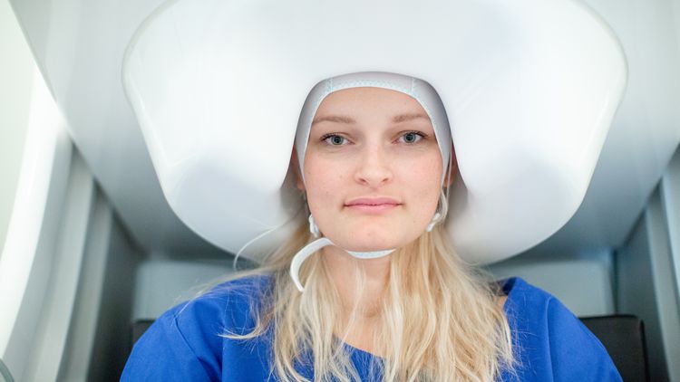
For the measurements to be carried out, the test person sits on a chair. Her head almost disappears under a device that resembles a huge version of the hood hair dryers at a hair salon. Under the white covering is a unit containing liquid helium cooled to a few degrees above absolute zero. The helium keeps approximately one hundred ultra-sensitive, superconductive sensors at the right operating temperature. Each sensor has three separate channels that measure the magnetic signals generated by nerve activity.
The MEG technique is used among other things to gain a better understanding of neurological diseases such as epilepsy and Parkinson’s. The Oldenburg researchers in Herrmann’s team, for example, are studying electrical brain stimulation – a technique that aims to either increase or decrease nerve cell activity in targeted areas of the brain.
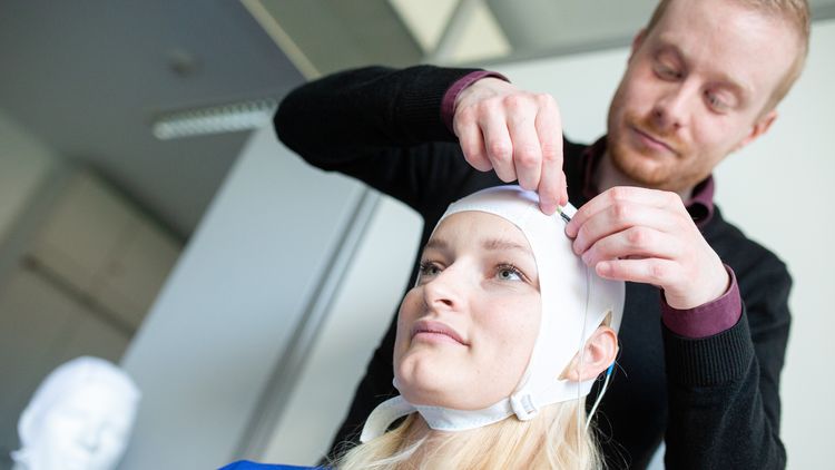
To do this, electrodes that generate low-frequency direct current or alternating current are attached to the scalp. A technique known as “Transcranial Alternating Current Stimulation” (tACS) can be used to influence the strength and frequency of electrical brain activity. In most cases the goal of the treatment is to normalise the activity of diseased regions of the brain. “So far, however, the effects of brain stimulation have for the most part been relatively weak and highly variable,” Kasten reports. Critics have therefore cast doubt on the effectiveness of the procedure as a whole.
Now the team of researchers led by Herrmann and Kasten has found a way to better predict the effects of the procedure on individual patients. “The result is a milestone on the path to future therapeutic applications of the technique,” says Herrmann. The researchers recently presented the results of their study in the scientific journal Nature Communications.
Variations in neuroanatomy play a key role
MEG played an important role in the investigations: the team studied the impact of individual differences in neuroanatomy on the effects of electrical brain stimulation. To do this, they first compiled structural images of the brains of 40 test persons using the University’s magnetic resonance imaging scanner. This enabled them to create maps of the individual electric field flow through the brain for each test person.
Twenty test persons then received a 20-minute session of brain stimulation, while the other twenty received a placebo stimulation treatment. Using the MEG technique, the researchers compiled detailed maps of the brain activity of the test persons before and after the session. The tests showed that brain stimulation had a strong effect when the computed map of electric fields in the brain created using MRI strongly resembled the brain activity map of a test person. When the similarities between the two weren’t as strong, the effects of brain stimulation were correspondingly weaker. These results mean that the researchers can now more accurately predict the effects of brain stimulation. Patients with disorders such as schizophrenia or Attention Deficit Hyperactivity Disorder (ADHD), for example, could benefit from the technique.


