Neuroimaging Unit
Neuroimaging Unit

The Neuroimaging Unit at the Carl von Ossietzky University of Oldenburg is a core facility of School VI – Medicine and Health Sciences at the University of Oldenburg and is located in the NeSSy research building on the Wechloy campus. Members of the Neuroimaging Unit come from various disciplines, such as psychology, medicine, and physics, and are part of School VI – Medicine and Health Sciences as well as the Cluster of Excellence “Hearing4all.”
The Neuroimaging Unit operates both a 3T magnetic resonance imaging scanner (MRI) and a magnetoencephalograph (MEG), which were both acquired exclusively for research purposes. These large-scale instruments were funded through the Cluster of Excellence, the University Medicine Oldenburg, and the German Research Foundation (DFG).
Equipped with various systems for auditory and visual stimulation, response boxes, eye tracking, and a self-developed driving simulator, we are able to measure and analyze sensory, functional, and cognitive functions of the brain and to gain new insights into these processes.
Magnet Resonance Imaging (MRI) scanner
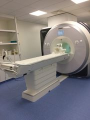
Siemens Magnetom Prisma (3 Tesla)
- 20 and 64 channel head coils, paediatrics head coil
- Headphones: OptoACTIVE with active noise cancellation and in-ear headphones Sensimetrics S14
- Response pads: Current Design with varios functions
- Projector: PROPixx, VPixx Technologies Inc.
- Eyetracker: Eyelink 1000
- custommade Driving simulator
- MR-compatible EEG: BrainProducts
- functional MRI (fMRI)
- structural MRI (T1 and T2 weighted)
- Resting State
- Diffusion (DTI)
- Spectroscopy (GABA)
- hMRI
Magnetencephalography (MEG) scanner
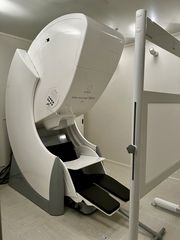
Elekta Neuromag Triux
- 306 channel MEG
- 102 triplet with 1 magneto- and 2 orthogonal gradiometers
- 128 channel EEG
- 12 bipolare biochannels
- TRACKPixx3 and SR Research EyeLink 1000 eye trackers
- driving simulator
- Polhemus FASTRAK digitizer
- 1440 fps PROPixx projector
- RESPONSEPixx handhelds with 1x4, 2x2, and 2x5 response buttons
- signal processing unit DATAPixx3


