Researchers at the University of Oldenburg are using electron microscopy images of SARS-CoV-2 to generate images that for the first time provide a highly detailed impression of the infection process. The new method relies on machine learning.
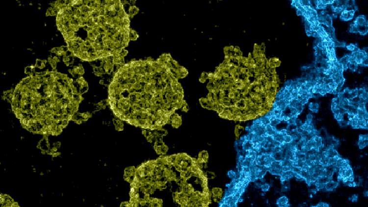
These days every child knows what a coronavirus looks like: something like a curled-up hedgehog with spikes that are wider at the top than at their base. However, many of the images of the virus currently in circulation are illustrations. To obtain a direct image of the virus itself, an electron microscope is needed. However, "Images of SARS-CoV-2 taken with these devices are quite blurred and appear flat or 2-dimensional," says Prof. Dr. Jörg Lücke, a machine learning expert at the University of Oldenburg's School of Medicine and Health Sciences. The microscopic images mostly show circular structures with small smudges on their periphery. The shape resembles a crown – in Latin: "corona" – which is how this group of viruses got its name.
Lücke is currently involved in a research project that studies methods for improving the quality of images of the pathogen. He and his doctoral students Jakob Drefs and Sebastian Salwig are focusing mainly on reducing the noise in electron microscopy images – or in other words, suppressing distorting statistical signals that have a negative impact on the quality of the images even at high resolution, making them look grainy. The three researchers are developing special algorithms that are based on various methods of machine learning, a key Artificial Intelligence (AI) technology.
Highly detailed images
Now the team has made its first major advances in the quest to get a clearer picture of the coronavirus. "We can calculate clear, denoised images from noisy electron microscopy close-ups of SARS-CoV-2," Lücke reports, adding: "We have also succeeded in generating a spatially appearing, high-resolution image on the basis of a single electron microscopy image." The team has published its preliminary results on a preprint server, so they have not yet been peer reviewed. The researchers are currently conducting further tests, and plan to present their work at an international conference in the near future.
As an example, one of the images shows SARS-CoV-2 viruses infecting a cell. In the colourised image, the pathogens look like spherical objects with irregularly shaped protrusions. These protrusions are called spike proteins. "However, they are not spiky, but rather rounded in shape, resembling little trees that lean in different directions," Lücke explains. The fact that these tiny structures are at all recognisable on an image that shows an entire infection scene testifies to how detailed the new images are, he adds.
The images also confirm the results of other research groups that have decoded the three-dimensional, molecular shape of the spike proteins on the basis of tomograms. Three-dimensional reconstructions of individual SARS-CoV-2 viruses already exist. However, unlike the Oldenburg researchers' images, they were reconstructed on the basis of a large number of electron microscopy images. According to the Oldenburg team, so far no other method has successfully produced images that give a spatial impression of an entire infection scene on the basis of a single electron microscopy image.
A new take on the opponent
The Oldenburg team's investigations are part of SPAplus, an interdisciplinary project coordinated by the Centre for Industrial Mathematics at the University of Bremen and funded by the Federal Ministry of Education and Research (BMBF) within the framework of its Mathematics for Innovations funding programme. Last year, the Oldenburg team received special funding from the BMBF to use its methods to improve the quality of images of coronavirus infection processes.
The new images could ultimately prove to have more than just scientific applications: Lücke hopes that they will also help to better convey the threat posed by these pathogens. "A 3D photo of a cell being attacked by coronaviruses which everyone can understand shows more vividly than an artificial model what kind of opponent we are dealing with," he says.


