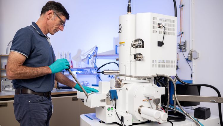

The University of Oldenburg has recently acquired a particularly powerful electron microscope, which is also available to external users. The high-resolution field emission scanning electron microscope can image structures as small as 0.5 billionths of a metre (nanometres).
The instrument is well-suited for working with sensitive materials, such as biological samples. It will be employed for measurements in a range of scientific disciplines, including physics, chemistry, biology, and medicine. The researchers plan to use the state-of-the-art microscope to study pollen and plant roots, elucidate metabolic processes in brain cells, characterise new catalyst materials, image the structure of nanomaterials and detect magnetic nanoparticles in biological cells.
For many of the research groups involved, it is crucial that the new electron microscope can be used to analyse wet or oily materials, such as biological tissue or plastics, with extremely high resolution. Two features of the instrument contribute to this. Firstly, unlike conventional electron microscopes, it is not necessary to apply conductive coatings to samples in order to examine them. Secondly, the investigations do not have to be carried out in an absolute vacuum; measurements can also be made in a low vacuum. However, it is also possible to image conductive samples in a high vacuum. This provides the highest possible resolution.
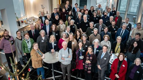
The total costs of the new microscope are around 800,000 euros. Half of this is being funded by the German Research Foundation (DFG) as part of its Major Research Instrumentation Programme, while the other half is being financed by eleven working groups at the Institutes of Chemistry, Physics, Biology and Environmental Sciences as well as the Faculty of Medicine and Health Sciences. The ultra-modern device is part of the Electron and Light Microscopy Service Unit at the Institute of Biology and Environmental Sciences, whose scientific director is Prof. Dr. Michael Winklhofer. Physicist Dr. Vita Solovyeva is responsible for the technical management. External interested parties can use the device at standard market prices.
This text has been translated from German with the assistance of DeepL.