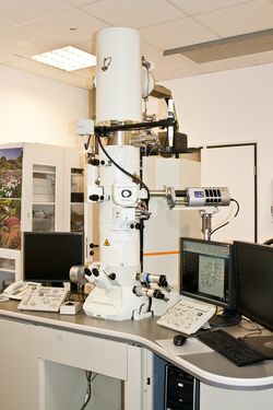
Jeol JEM2100F TEM
Kontakt
Scientific director
Technical Director
Team
Manuals
Jeol JEM2100F TEM
 The Jeol 2100F electron microscope is a high resolution scanning/transmission electron microscope equipped with Schottky field emission gun (FEG). It is well suited for material sciences, and can be operated at voltages between 80-200 kV in both, the transmission (TEM) mode and the scanning transmission (STEM) mode.
Additionally, it offers chemical analysis and tomography capabilities. It is well suited for high-resolution imaging in the TEM mode, and atomic resolution can be achieved due to the high resolution objective lens polepiece. Z-contrast imaging is possible in the STEM mode using the bright field (BF) and high angle annular dark field (HAADF) STEM detectors. Specimen tilts of ±30° are possible with the regular sample holder, tilts up to approximately ±80° with a high tilt holder. An Oxford energy dispersive X-ray analysis system allows elemental analysis. The high tilt holder in combination with the Tomography software allows 3D-rendering of samples. Digitized image documentation is achieved via two retractable CCD cameras.
Specifications Jeol 2100F S-TEM | |
| Accelerating voltage | 80 kV - 200 kV |
| Magnification | 50x – 1,500,000x |
| Magnification steps | 30 steps in Mag Mode (2,000x - 1,500,000x) 20 steps in Low Mag Mode (50x - 6,000x) 21 steps in SA Mag Mode (8,000x - 800,000x) |
| Resolution | 0.14 nm lattice image 0.23 nm point image |
| Camera length steps | 15 steps in SA DIFF (80-2,000mm) 14 steps in HD DIFF (4-80mm) 1 step in HR DIFF (333mm) |
| Objective lens polepiece | high resolution polepiece (HRP) |
| Focal length | 2.3mm |
| Spherical aberration coefficient | 1.0mm |
| Chromatic aberration coefficient | 1.4mm |
| Minimum focal step | 1.5nm |
| Exciting current stability | 1 ppm/min. |
| Specimen stage | micro active goniometer |
| Specimen tilt angle | ±35° (X-axis); ±30° (Y-axis) |
| Specimen movements | 2.0mm (X); 2.0mm (Y); 0.4mm (Z) // (±0.2mm) |
| Additional equipment | |
| EDX | Oxford AZTEC with SDD-Detector X-Max80 (area 80 mm2, resolution 129 eV at Mn K; detects elements from Be to Pu) |
| Tomography | High tilt specimen retainer; Jeol Tomography software packages for acquisition, 3D-reconstruction, and 3D-rendering |
| Image documentation | above the viewing screen: Gatan Orius SC200D (2k x 2k; 15mm x15mm, side entry lens coupled camera; readout of up to 30fps) below the viewing screen: Gatan Orius SC600 (2.7k x 2.7k; 24mm x24mm, fiber optic coupled camera; readout of up to 13fps) operating software: Gatan Digital Micrograph |
