
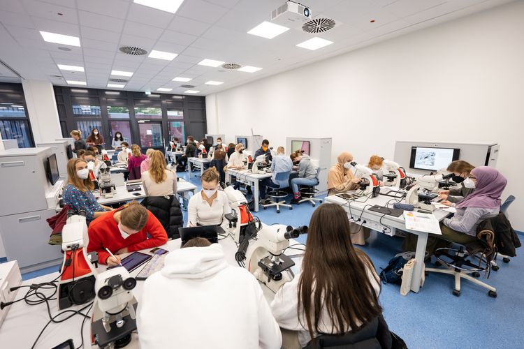


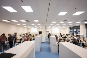
A new 200 square metre microscopy room* has been created in the former carpentry and metalworking rooms on the ground floor of the A03 building on the Haarentor campus of the University of Oldenburg. So that it can be used for different courses, it can be flexibly divided into two roughly identical individual rooms using a partition wall.
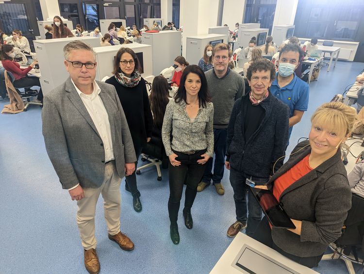
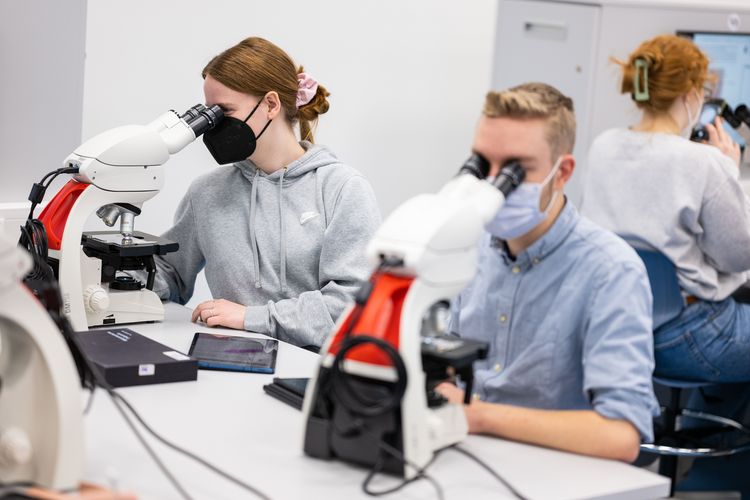
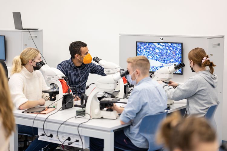
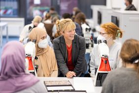
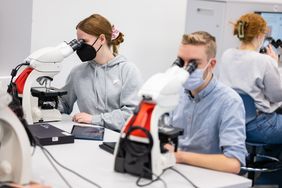
The microscopy room has space for 64 students, who can work here with Leica ICC50 W light microscopes. A special feature: they can look at their specimens together with their fellow students and lecturers and discuss the image together. The lecture theatre is designed so that students can sit and work together in groups of four. Each table of four has a monitor on which the image of a microscope can be transmitted. "This means that the concept of small group teaching can be optimally implemented in the model degree programme in human medicine despite the large number of students," says anatomist Prof. Dr Anja Bräuer, who played a key role in the room concept and held the first course in the new room. Many ideas came together, for example the tables and cupboards were all produced in-house. They were designed by project manager Ajana Milanovic from Division 4 and made by the university's carpentry department.
"The design of the new microscopy room is unique in Germany. We now have a state-of-the-art, well-equipped microscopy room in which the images from the microscopes can be streamed to the respective monitors and also to the projection screen behind the lecturer's desk. This is a great support for our teaching," says Dean of Studies Prof Dr Karsten Witt.
The entire remodelling took one year. Due to the increase to 120 students in human medicine from the winter semester 2022/23, Division 4 had to complete the construction work on a tight schedule by October 2022 in order to ensure that microscopy courses could be taught. In addition to human medicine, the new microscopy room is also used by other degree programmes such as chemistry, physics, biology or the Oldenburg Teaching-Learning Rooms (OLELA) as a theory-practice room.
*With an additional seminar room, corridor and storage area, the total conversion area was around 340 square metres.
Further information at: