
The University's Department of Psychology is regularly looking for test subjects for various studies. What can the test subjects expect? Our editor Ute Kehse tried it out: she took part in a study on chronic pain.
Before we start, Melanie Spindler asks me lots of questions: Do I really not have any metal anywhere in my body - for example, an artificial joint, the remains of braces or a tattoo? Have I ever had an epileptic seizure? What about migraines? She asks me to remove any coins or mobile phones from my trouser pockets and take off my belt.
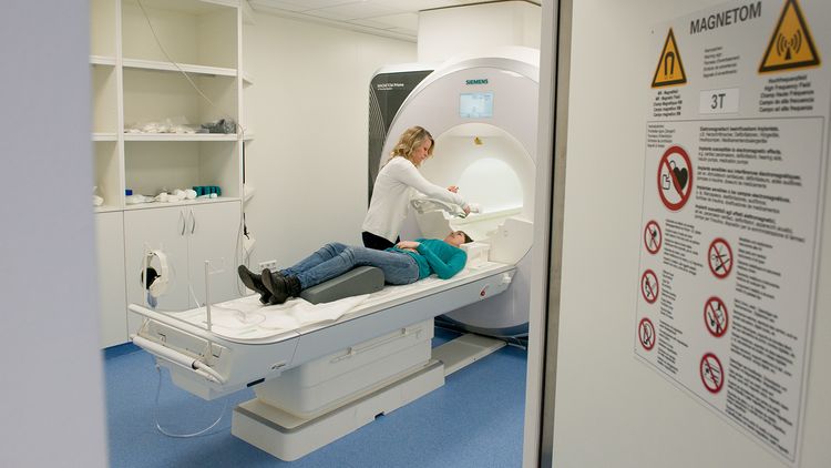
These precautions are necessary because Spindler and her colleague Gülsen Yanc are about to push me into a white monster in the next room - the university's magnetic resonance imaging (MRI) scanner, which has been used primarily in neuropsychological, audiological and medical research since 2016. The device, which weighs several tonnes, generates a strong magnetic field and could, for example, set metal parts in the body in motion. Finally, Spindler asks the most important question: do I suffer from claustrophobia? Not really, but the sight of the narrow tube does make me feel a little uneasy. Gülsen Yanc reassures me: "If I feel uncomfortable, I can press a grade button at any time. "Then we'll stop the measurement and get you out," she says. "The most important thing is that you feel well."
Does chronic pain change the brain?
The patient couch is surprisingly comfortable, with a cushion under the legs it is quite relaxed. Yanc, an X-ray technician in the Biological Psychology Laboratory, attaches a white frame over my head. "This is a coil that we need for the measurements," she explains. The "ChroPain2" study I'm taking part in is about chronic pain. It is a project led by Dr Carsten Bantel, Senior Physician at the University Clinic for Anaesthesiology and Pain Therapy in co-operation with Prof. Dr Christiane Thiel, Head of the Department of Biological Psychology. Melanie Spindler, a student on the Neurocognitive Psychology Master's degree programme, is supervising the measurements. The team wants to find out whether the constant joint pain suffered by rheumatism patients alters certain areas of the brain. There are indications of this: "Some studies show that chronic pain affects cognitive processes such as attention," reports Spindler. This seems plausible: who can concentrate well if you are constantly hurting somewhere? Other MRI examinations have also shown that the structure of the brain changes in pain patients.
I belong to the healthy control group: The research team is trying to find a healthy person of the same age and gender for each patient taking part in the study. In this way, the researchers want to find out whether the brains of sick and healthy people actually work differently.
Still in the dark
I now have to go into the tube for this. Because the tomograph rumbles as loudly as a jet plane taking off during measurements, I am given earplugs - and told to move my head as little as possible. Just at that moment, I am overcome by the urge to cough. What should I do if this happens during a measurement? "It would be great if you waited to cough until the device is not measuring," says Yanc. "If you move your head more than three millimetres, the measurement will be unusable." All right, I'll give it a try. The couch slides into the round opening of the device. I look through a mirror at a screen inside the MRI tube.
I quickly forget the confinement around me. "You can close your eyes now and don't need to do anything else," says Yanc, who is now sitting in the neighbouring room and gives me instructions via a microphone. The screen goes dark and the measurement begins. A muffled humming sound lasting perhaps two seconds repeats itself continuously. The noise is caused by coils in the wall of the MRI scanner. They generate the magnetic field for the measurement and are set into oscillation by strong alternating currents. For six minutes, the device takes images of my brain layer by layer. "We need these high-resolution images as the basis for our study," explained Melanie Spindler in the preliminary discussion. At first, the time seems incredibly long. How am I supposed to last an hour in here without moving? But then my thoughts drift off despite the droning noises and I relax.
Suddenly it gets light again and Gülsen Yanc announces that the first real task is about to begin. Now it's getting a bit stressful: A subtraction task appears on the screen: 98 - 13. Melanie Spindler has explained to me that I should say the result of the maths task loudly and clearly and then keep subtracting the subtrahend from the result. So I speak against the sound of the MRI, which now sounds like a mixture of a jackhammer and a dentist's drill. 85 - 72 - 59 - 46... The time is up and the programme continues with a new task. Then the programme gives me a little break: I have to fixate on a small, bright cross on the black screen for a few moments until the next calculation task appears.
Grey cells in action
The programme alternates between calculations and short breaks for seven minutes, followed by another measurement cycle. This is followed by a new task: this time I have to estimate distances on a line. With these tasks, the researchers are not only interested in the differences between pain patients and the control group, Melanie Spindler explains to me later. She and her colleagues also want to use MRI to measure the activity of certain brain regions. They are particularly interested in the "intraparietal sulcus", a groove in the parietal lobe of the cerebrum. "It is assumed that this is where number processing takes place," explains Spindler. In order for this region to become active, the grey cells there have to be given something to do.
Finally, the jackhammer noises disappear and it becomes light again in the tube. I am glad that I survived the MRI examination without a coughing fit. Now we move on to a neighbouring room, where Melanie Spindler has more tasks on the agenda, this time without high-tech equipment. "For example, we measure spatial and verbal working memory and the processing speed of the brain. We also carry out a small intelligence screening," she explains.
In an earlier study, the Oldenburg researchers had already discovered that pain patients were worse at estimating distances on the number line than the control group. The team has just published the results in the journal Frontiers of Behavioural Neurosciences. "We want to use the MRI measurements to find out where this difference comes from - for example, whether different areas of the brain are activated in pain patients than in healthy test subjects," she explains.
Help for pain patients
The study should help to better help patients with chronic pain. "Pain patients very often have to assess their pain intensity during examinations, usually using a visual analogue scale that resembles a number line," says Spindler. The team's reasoning: if the number sense of people with chronic pain is actually impaired, this method may not be as suitable. They now hope to find new ways to assess the intensity of the pain more accurately.
For me, the examination is now over. The two and a half hours have flown by. For Melanie Spindler, however, the work is only just beginning: she has to analyse the results of the MRI calculations, the MRI images and the results of the tests. It will therefore be a few months before the study results are available. Spindler promises: "As soon as there is a publication, she will inform me by email. I'm excited - and hope that the results will actually benefit pain patients soon.