For experts
For experts
Attosecond microscopy on surfaces
We combine photoemission electron microscopy (PEEM) with XUV light, which we generate as the high harmonics of our femtosecond laser pulses. We achieve photon energies of up to 140 eV in argon and potentially significantly higher energies in neon and helium. We focus these pulses onto a sample surface using a toroidal mirror or dielectric mirrors. We analyse the emitted electrons using time-of-flight photoemission electron microscopy. Our setup is particularly characterised by the fact that we can focus the XUV pulses onto the sample with a focal length of 125 mm and the IR pulses with a focal length of 33 mm. This allows us to achieve particularly high intensities in a small area.
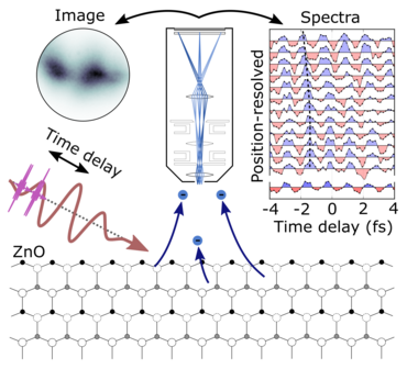
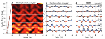
The precursor experiment from our setup in Lund was published in Advanced Physics Research at the end of 2023, shortly after the Nobel Prize was awarded to attosecond research. In it, we were able to perform a time-resolved experiment with attosecond pulses in a PEEM for the first time. For this purpose, we used XUV-induced electron emission from a freshly prepared ZnO surface. A time-delayed few-cycle pulse around 800nm provided the transient electric field strength to periodically accelerate the emitted electrons. We were able to demonstrate this by means of a retarding field analyser in the PEEM and thus measure the spatiotemporal differences between the carrier frequency at 800 nm and the XUV pulses directly on the ZnO surface.
The findings from this experiment have been incorporated into our experimental setup in Oldenburg and we are currently working on the next steps.
BIRDS pulse duration measurement method
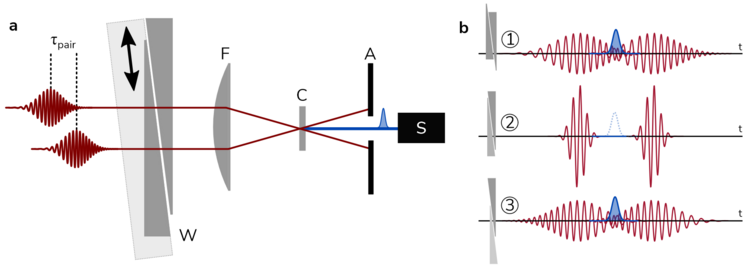
We have developed a new, relatively specialised measurement method for determining a pulse duration: "bi-pulse reconstruction via dispersion scan", or BIRDS for short. It uses an interferometer like in a FROG setup, but then changes the dispersion of the generated pulse pair like in a dispersion scan setup. The particularly exciting thing about this method is that no measurement signal is generated with optimum pulse compression. This only arises when a certain amount of dispersion (with both signs) has been added to the pulse pair. In this way, the method is particularly sensitive to pre- and post-pulses, e.g. caused by higher orders of dispersion. We have extended the COPRA algorithm with a reconstruction option for our measurement data and published the results in the journal Optics Express.
Femtosecond dynamics in PEEM
Even without attosecond pulses, exciting charge carrier dynamics in nanostructures can be investigated using photoemission electron microscopy. For this purpose, we use spectrally tunable few-cycle pulses in the visible to near-infrared spectral range. A first pulse excites charge carriers in the sample to higher energy levels, where they begin to develop their dynamics. After a variable waiting period, a second pulse drives the photoemission of the charge carriers. These carry valuable information about the dynamic state of the sample. We therefore analyse the emission location, energy (spectrum) and angular distribution of each electron with our experimental set-up. For example, we learn how charge carriers behave after optical excitation in InAs nanowires, i.e. specifically which relaxation processes play a role on which time scales and how spatially inhomogeneous field distributions influence the dynamics. Not only nanostructures, but also 2D materials are of particular interest, as they can also become relevant for optoelectronic assemblies and therefore fundamental processes of light-matter interaction must be understood. The interfaces occurring in such devices are particularly important, as this is where the macroscopic function is defined. Our spatiotemporal resolution allows us to investigate the dynamics precisely there. In parallel, we are expanding our experimental setup to include additional light sources, in particular an HHG beamline with a single, spectrally narrow harmonic for photoemission.
