
Radiotherapy is one of the three pillars of cancer treatment, alongside surgery and drug therapies. Oldenburg scientists have been researching measuring devices that record the distribution of radiation doses in the body for ten years. A contribution by medical physicist Björn Poppe and the "Medical Radiation Physics" working group.
Radiotherapy is always a balancing act: it must dose the high-energy ionising radiation in such a way that as much tumour tissue as possible is destroyed and healthy tissue is spared. This requires precise information about the tumour and its metabolism. This information is obtained using complex mathematical calculations and various imaging techniques. It is usually medical physicists who plan the radiotherapy and work in close partnership with the doctors. They have developed methods with which the distribution of the respective radiation dose can be calculated in advance - in a three-dimensional, computerised tomographic model of the body. This method is now standard in all radiotherapy.
The aim: to significantly reduce the side effects of radiotherapy
All types of radiation that are able to penetrate the body and deposit part of their energy there by ionising atoms are suitable for therapy. Ionisation is a process in which an electron is ejected from an atom and the atom remains as a positively charged ion. In this way, the atoms acquire new properties that can lead to the breaking of molecular bonds in the cell and to severe damage to the DNA - which can cause the cells to die. In order for the healthy tissue to recover better, the entire radiation dose is divided into small portions, so-called fractions, with which the patients are irradiated in numerous individual sessions. Ideally, the tumour shrinks steadily until it dies off completely. The problem remains the reaction of the healthy tissue and the associated side effects. They set the limits for the maximum radiation dose that can be administered.
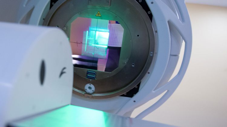
This is where the linear accelerator comes into play, without which modern cancer therapy would be inconceivable. It accelerates electrons until they build up high energies and then slows them down abruptly. Part of the kinetic energy is converted into high-energy X-rays, which are then directed at the patient. To ensure that as little healthy tissue as possible is damaged, a lamella collimator attached to the linear accelerator adjusts the beam to the tumour. This literally "cross-fires" the tumour by rotating the radiation arm from different directions.
In recent years, intensity-modulated radiotherapy (IMRT) has become established in practice. It uses lamella collimators not only to block out organs at risk, but also to change the radiation intensity. The lamellae either move continuously over the area to be irradiated or the beams act on it from several directions in various field configurations. In the latest devices, the irradiation arm and the lamellae rotate dynamically so that almost any rotationally symmetrical dose distribution in the body can be realised.
"Medical Radiation Physics" working group"
The new methods have significantly reduced the massive side effects of radiotherapy that were still common twenty years ago. Nevertheless, they are still at the limit of what can be tolerated by healthy tissue. Even the slightest inaccuracy in dosage can cause completely different side effects in patients. However, it was difficult to measure and check the dose distribution - which is why many clinics did not use intensity-modulated radiotherapy for a long time.
The Oldenburg "Medical Radiation Physics" working group, which is jointly supported by the University of Oldenburg and Pius Hospital, is working on developing highly accurate measuring devices that record the dose distribution in the body using intensity-modulated radiotherapy. They rely on measuring the radiation dose in body-like materials such as water or plastics, in which they place detectors.
Before intensity-modulated radiotherapy, it was mainly simple flat and homogeneous intensity profiles that were irradiated into the human body. Measurements were generally limited to point detectors. The standard measuring device for this is the ionisation chamber - an air-filled cavity in which the voltage of two electrodes generates an electric field. The electrons generated by the radiation are attracted to the electrodes, where they generate a signal that provides information about the radiation dose.
Advances in irradiation techniques require measurement techniques that can record more complex dose distributions. Initially, there was no experience as to whether the mathematical methods developed for relatively simple field shapes could be transferred to the complex and dynamic techniques of IMRT. Precise, multi-dimensional measurements are required to compare the calculated dose distribution with the actual dose distribution in the body.
Developing two-dimensional detectors based on ionisation chambers
X-ray films were usually used for this purpose. However, research by the Oldenburg working group showed that they no longer meet the increased demands because they do not adequately depict the end position of the radiation in the body. Added to this was the digitalisation of X-ray technology. It has replaced conventional X-ray film and the associated developing machines. It was therefore necessary to develop new detectors that took account of the advances in radiation research. The digital detectors initially used in radiology are also only of limited use. On the one hand, the radiation energy is so high that the devices, which are usually CCD-based, are destroyed relatively quickly. On the other hand, the physical properties of the detectors mean that precise dose measurements can only be carried out at extremely high cost.
As an alternative, the Oldenburg physicists, together with scientists from the Freiburg Physikalisch-Technische Werkstätten (PTW), have developed two-dimensional detectors based on ionisation chambers. In these detectors, the ionisation chambers are arranged in one plane - similar to pixels in a digital camera. The detectors are significantly larger than the individual pixels of a CCD chip, for example. But how large can the detectors be and how many are needed for a precise comparison between the predicted and actual dose distribution?
The physicists found the answer in multidimensional signal processing. In order to put this method into practice, they first had to adapt the mathematical description of the dose deposition and apply it to the measurement methods. This was the only way they could estimate the necessary and optimum number of chambers and their size. For practical use, they first realised a detector with around 1000 measuring chambers. This allows the dose distribution to be checked with sufficient accuracy for most clinical applications.
After initial scepticism among physicists and doctors, the design of these chamber arrays has now become established worldwide. In recent years, other research groups have also confirmed the results of the Oldenburg and Freiburg scientists and developed detectors based on their principle. Today, every radiotherapy centre that offers intensity-modulated techniques is likely to have such a detector array.
Ion beams allow more precise irradiation
But that was just the beginning: theoretical analyses of radiation transport in the human body have shown that there is a minimum value for the detector size and the distance to the patient. This is in the range of two and a half millimetres in each case. Below this, no improvement can be achieved - even theoretically - due to the interaction between radiation and matter with the photon beams normally used. In practice, the inaccuracies increase and are in the region of around five millimetres. Ion beams could enable even more precise irradiation. However, their research is still in its infancy and is a long way from routine clinical use.
The Oldenburg scientists and their partners are therefore focussing on a detector array that works at the minimum resolution limit. Air is no longer an option as a detection medium due to the small volume of the chambers. This is replaced by electrically non-conductive liquids, such as isooctane. The different physical properties of the liquids pose new challenges for medical physics. For example, the number of chambers in the devices of the future must increase to well over 1000 - which in turn requires a signal-theoretical optimisation of the arrangement on the array.
As part of a co-operation with partners from Ashland Inc. in Wayne, New Jersey (USA), the Oldenburg scientists are also researching monomers. This type of molecule could optimise ionising radiation for dosimetry due to its chemical reaction. After absorbing the radiation, the monomers form polymers that have a different light absorption. If these monomers are applied to a thin film-like base, the altered light absorption results in a kind of "blackening". As the size of these monomers is only in the range of a few micrometres, it is in principle possible to increase the resolution of the measurements almost at will. The physical properties of these processes and their dosimetric applicability are the subject of various worldwide investigations involving Oldenburg Medical Physics.
The ultimate aim of all these efforts is to optimise the match between calculated and applied dose in order to further improve radiotherapeutic techniques. To ensure this, physicists and physicians work together more closely in radiotherapy than probably in any other area of modern medicine.
The article appeared in the current issue of EINBLICKE. The authors are Björn Poppe, Kay C. Willborn, Hui Khee Looe, Ndimofor Chofor.