Methoden und Geräte
Methoden und Geräte
Molekularbiologie
Geräte | Techniken/Methoden |
Eppendorf Mastercycler nexus Gradient | PCR |
|
|
|
|
|
|
|
|
|
|
|
|
Lipidbiochemie
Geräte
CAMAG TLC Sampler 4 CAMAG TLC Visualizer CAMAG TLC Scanner CAMAG Derivatizer | Techniken/Methoden
HPTLC von Lipiden |
|
|
Proteinbiochemie / Histologie
Geräte Beckman Coulter Optima max-XP Ultrazentrifuge mit Rotor MLS-50 (Swinging-Bucket-Rotor) und Rotor TLA-110 (Fixed Angle Rotor) | Techniken/Methoden Proteinfraktionierung |
|
|
|
|
|
|
|
|
|
|
Geräteraum
Geräte New Brunswick Innova C585 Ultra | Techniken/Methoden |
|
|
|
|
|
|
|
|
Taylor Wharton LS-3000 Stickstoff Vorratsbehälter |
Zellkultur
Geräte 2x New Brunswick CO2 Inkubator Galaxy 170S | Techniken / Methoden Kultivierung prim. Neurone, Astrozyten, HEK-Zellen, Hap1-Zellen |
|
|
|
|
|
|
Olympus CKX53 : inverse Phasenkontrast Technologie | Mikroskopie von Zellkulturproben |
Mikroskopie
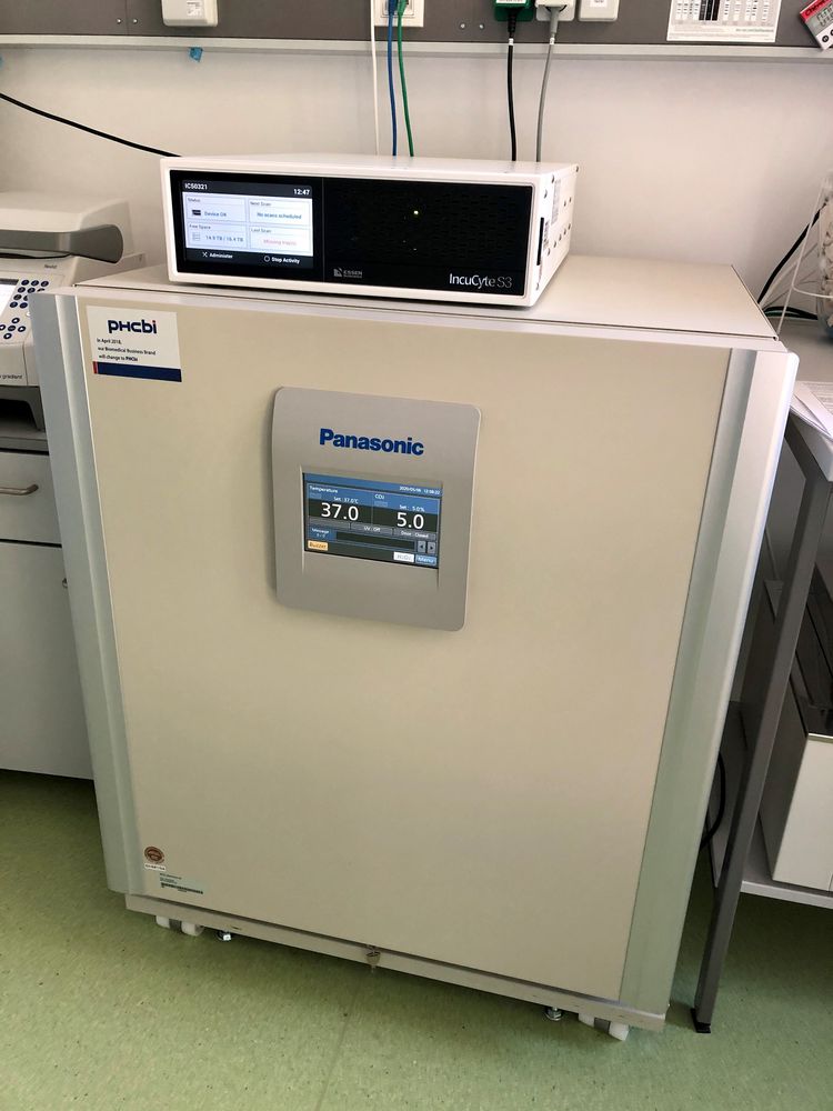
IncuCyte S3 _ Live-Cell Imaging and Analysis
Objectives: 4x, 10x, 20x, air
Excitation/emission: 440-480nm/504-544nm (Green) and 565-605nm/625-705nm (Red).
Temperature regime: 0-42C.
CO2 Incubator
The image analysis software provided by IncuCyte is also available for the users.
Live-Cell Analysis System Applications: Software Modules
- Basic Analyzer (Stem Cell Monitoring and Reprogramming, Cell Culture QC, Dilution cloning, Transfecion Efficiency, Reporter Genes, Proliferation, Apoptosis, Cytotoxity, Immune Cell Clustering, Antibody Internalization, Immune Cell Killing, Phagocytosis)
- Neuro Track (Neurite Analysis)
Incucyte Vessel Trays available for: 35 mm dishes, T25 flasks, 6-/12-/24-/96-well plates
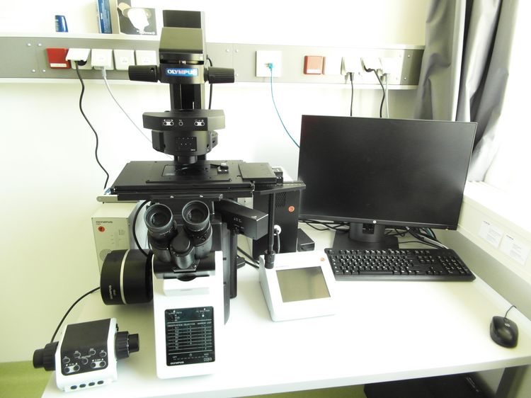
Olympus IX 83 inverses Fluoreszenzmikroskop
Lichtquelle:
Cool LED pE-4000 (4-channel high-specification LED System mit 16 LEDS, 365-770nm)
Objektive:
- UPlanFLN 4x/0.13 PhL
- UPlanSApo 10x/0.4
- UPlanSApo 20x/0.75
- UPlanSApo 40x/0.95
- UPlanSApo 60x/1.35 Oil
- UPlanSApo 100x/1.4 Oil
Filtermodule:
- U-F39002 AT-FITC (Acridine Orange, Alexa Fluor488, eGFP, Fluo 4, FITC, Oregon Green 488)
- U-F39003 AT-EYFP N/C (eYFP)
- U-F39004 AT-CY3 (Alexa Fluor 546, Alexa Fluor 555, Cy3, Dil, TRITC)
- U-F39007 AT-CY5/ALFL647 (Alexa Fluor 647, APC, Cy5, TO PRO 3)
- IX3-FDICT (Differentialinterferenzkontrast, DIC)
- U-FF (DAPI)
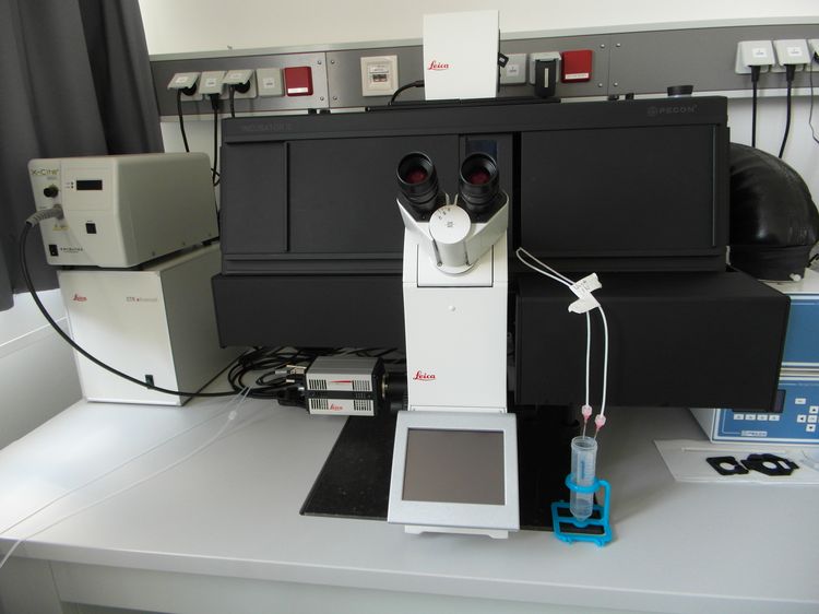
Leica DMi8: inverses teilmotorisiertes Forschungsmikroskop mit Ca-Imaging Ausstattung
Objektive:
HC PL FLUOTAR L 20x/0.40 Dry (LMD; PH1)
HC PL FLUOTAR L 40x/0.60 Dry
HC PL FLUOTAR 40x/1.30 Oil (Fura)
HC PL APO 63x/1.40 Oil
HC FLUOTAR 100x/1.30 Oil (Fura)
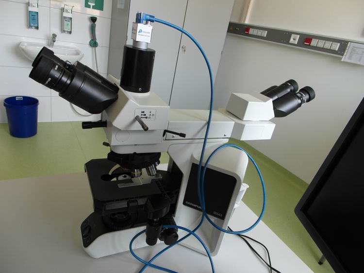
Olympus BX43F aufrechtes Durchlichtmikroskop mit Digitale Hochleistungs-Mikroskop-Kamera für Scannen
Lichtquelle:
True Colour LED (Farbwiedergabeindex (>96), Intensität äquivalent mit 30 W Halogen)
Objektive:
6-fach Objektivrevolver mit
- PLCN 4x/0.1
- PLCN 10x/0.25
- PLCN 20x/0.4
- PLCN 40x/0.65
- UPlanSApo 20x/0.75
Einheit für digitales Scannen:
- Basler acA2440-Kamera mit hoher Bildwiederholungsrate in Echtzeit 5.0 MP, mit Sony IMX264 Sensor Bildwiederholungsrate: 35 fps USB 3.0
- EasyScan Software, zum manuellen hochauflösenden Scannen von histologischen Präparaten
Mitbeobachtermodul:
Zweifach-Beobachtungstubus mit beweglichem LED Markierungspfeil
